
Fractures during clinical function have been reported as one of the major causes for failure associated with all-ceramic dental restorations. Comparison of fracture strength and fracture modes of different all-ceramic crown systems is not straightforward. Established methods for reliable testing of all-ceramic crowns are not currently available.
Previously, researchers have used a method in which a steel ball has been loaded to the occlusal surface of crowns. The fractures observed in these studies have not shown fracture morphology similar to clinical fractures in all-ceramic crowns. The cracks observed started on the occlusal surface while clinical fractures often occur at the cervical margin in approximal areas. An explanation could be that the steel ball may have been too tough for the ceramic surface and resulted in contact damages initiating the fractures.
 A new method for simulation of clinical fractures in ceramic crowns was recently presented (see Dental Materials 2013; 29:815–823). A rubber cushion is placed between the crown and the steel ball to avoid contact damages and a yielding material (epoxy) is used for abutments which result in strain at the cervical margin. This strain results in detrimental tensile forces to the crown and cracks occur at the cervical margins at a lower load than expected, as for the clinical ones. The method was evaluated using three different all-ceramic crown materials.
Analysis of ceramic crowns fractured in situ has been further elaborated (see Eur J Oral Sci 2014: 122: 238–244). This study gives a better understanding of the fracture start and propagation in a clinical setting.
Using the recently presented in vitro method, three all-ceramic systems were compared. Ten incisor crowns of three types of all-ceramic systems were exposed to soft loading until fracture. The initiation and propagation of cracks in these crowns were compared with those of a reference group of crowns that failed during clinical use. All crowns fractured in a manner similar to fracture of the clinical reference crowns. The present in-vitro method is well suited for evaluation of clinical fracture morphologies, relevant for different all-ceramic materials.
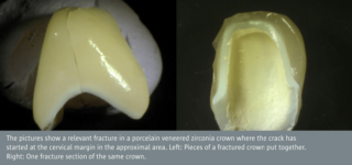
The zirconia crowns fractured at statistically significantly higher loads than alumina and glass-ceramic crowns. Fracture initiation was in the core material, cervical in the approximal areas. The pictures show a relevant fracture in a porcelain-veneered zirconia crown where the crack has started at the cervical margin in the approximal area.
The full study is described in the following
Øilo M, Kvam K, Gjerdet NR.
Simulation of clinical fractures for three different all-ceramic crowns.
Eur J Oral Sci 2014: 122: 245–250.
Download
NIOM NewsletterAugust 2014