
New scanning electron microscope
The new table scanning electron microscope can make 3D-reconstructions of a surface.
The SEM images are usually in black and white, but can also, per request, be coloured.

NIOM have 17 laboratories, and a vast, up- to date instrument park. Today we would like to highlight our new key instrument in materials research, nicknamed the SEM.
– Our new table scanning electron microscope (SEM TM4000Plus) is a crucial tool in our research activity. With this, we can evaluate the surface of many different materials. It can both image and analyse the elemental composition of the surface, NIOM Scientist Amund Ruud, Ph.D., explains.
An additional advantage is the possibility to make 3D-reconstructions of a surface. This gives the researchers important information about samples profile and roughness.
Practical use
In one ongoing project, the SEM is used to explore wear on dental implants.
-We can see tiny fractures and identify possible weak spots, Ruud says.
A guest researcher, assisted by Ruud, conducts the project.
– To me it is all about knowing techniques, and using the right tool for each research project. The SEM is both useful and versatile, Ruud says.
For those interested at conducting research at NIOM, as a guest researcher, or by renting time, it is possible to include time with our SEM operator.
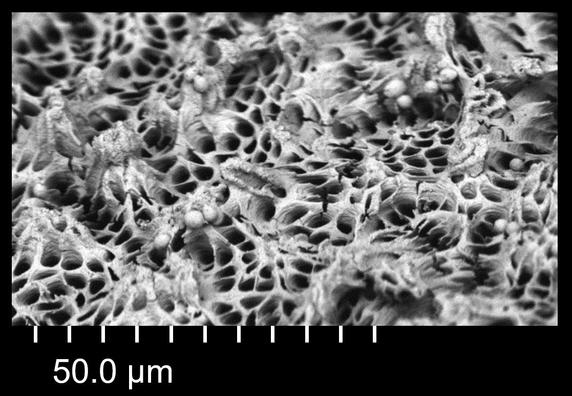
Credit: Amund Ruud. Image of cross section of a tooth, by the new SEM.
NIOM Newsletter January 2020